중심부 석회화를 동반한 간내 담관암 1예
요약
중심부 석회화를 동반한 간내 담관암은 국외에서 1예 보고되었을 정도로 매우 드문 증례이며, 담관내 석회화 소견을 보이는 환자의 감별진단에 있어서 간내 담도암에 대한 고려와 조직검사가 필요할 것으로 판단되어 중심부 석회화가 동반된 간내 담관암 증례를 보고하는 바이다.
Keywords: 담관암, 간내, 중심부, 석회화
Abstract
A 50-year-old woman complained of jaundice and dyspepsia that started 2 weeks prior to consultation. Abdomen-pelvic computed tomography showed a 3 cm mass in the right hepatic duct with central calcification, which was spreading into the second branch. Repeated biopsies through endoscopic retrograde cholangiopancreatography were needed for pathology, which was consistent with an adenocarcinoma. Imaging studies including positron emission tomography showed no evidence of distant metastasis. The patient underwent right lobectomy with bile duct resection. The final diagnosis was intrahepatic cholangiocarcinoma with central calcification. We reported a very rare case of centrally calcified mass growing in the second branch of the right hepatic duct. The possibility of intrahepatic cholangiocarcinoma with central calcification should be considered for differential diagnosis of intrahepatic calcification.
Keywords: Cholangiocarcinoma, Intrahepatic, Central, Calcification
서 론
담관암은 담관의 상피세포에서 기원한 악성 신생물로 정의되며 그 발생 빈도는 10만 명당 1-2명으로 드문 암이지만[ 1], 간에서 발생하는 암 중 간세포암종 다음으로 두 번째로 많은 비중을 차지하며 그 발생빈도는 꾸준히 증가하고 있는 추세이다[ 2]. 담관암은 위치에 따라 간내 담관암, 간외 담관암 그리고 클라스킨 종양(Klatskin tumor)으로 분류될 수 있으며 담관암의 전형적인 조영증강 computed tomography (CT)의 소견은 지연기에서 조영되는 담관 내부의 종괴 소견이며 전체 간내 담관암의 15% 정도에서 종괴의 석회화를 보일 수 있다. 대부분의 경우 종괴 변연부의 석회화를 보이게 되며 중심부의 석회화를 보이는 경우는 국외에서 하나의 증례보고가 있었을 뿐[ 3] 국내에서는 아직 보고된 바가 없을 정도로 매우 드물다. 게다가 간내 석회화는 간결핵, 문맥혈전증, 간혈관종, 간세포암, 기생충감염, 다른 암의 간전이, 간샘종 등 다양한 질환들에서도 동반될 수 있어 영상검사에서 간내 석회화가 발견되었을 경우 간내 담관암을 감별진단하기에는 어려움이 따른다[ 4- 7]. 최근 본원에서 중심부 석회화를 동반한 간내 담관암을 진단하여 수술적 치료를 시행하게 된 증례를 경험하였기에 보고하는 바이다.
증 례
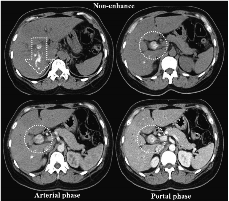
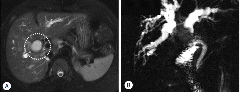
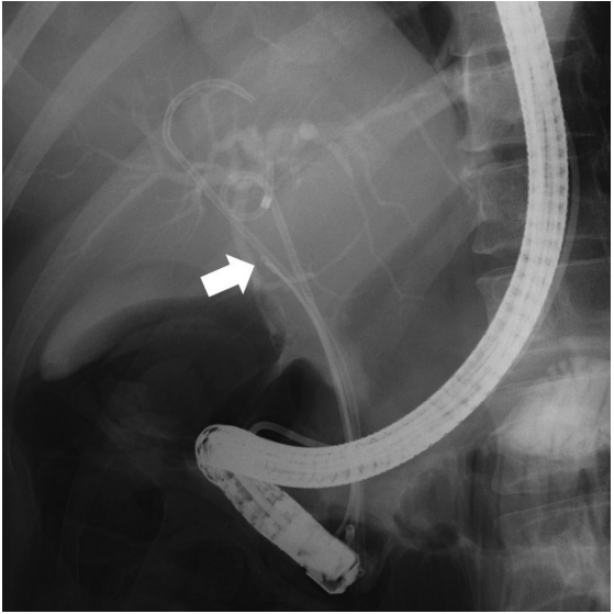
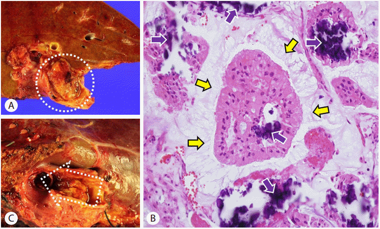
50세 여자 환자로 2주 전부터 황달과 소화불량이 지속되어 개인의원을 내원하였고 간내 담석과 담관염이 의심되어 정밀검사를 위해 본원으로 전원되었다. 30년 전 환자는 자궁경부암으로 방사선 치료를 받았던 과거력이 있었고 고혈압으로 혈압약을 복용 중이었으며 흡연력이나 음주력은 없었다. 입원 당시 활력징후는 혈압 140/100 mmHg, 맥박수 82회/분, 호흡수 20회/분, 체온 36.0°C로 측정되었고, 신체검사에서 공막황달과 우상복부에 압통은 있었으나 머피징후(Murphy’s sign)는 없었다. 혈액검사에서 백혈구 4,890/mm 3, 혈색소 12.8 g/dL, 혈소판 378,000/mm 3였고, 총빌리루빈은 7.08 mg/dL, 직접빌리루빈은 5.54 mg/dL, AST는 55 IU/L, ALT는 116 IU/L, ALP는 229 IU/L, GGT는 310 IU/L였다. 그리고 종양표지자 중 CA 19-9가 56.1 U/mL로 증가되어 있었다. 복부 CT 검사 결과 양측 간내 담관의 확장과 우측 담관내에 약 3 cm 크기의 경계가 불규칙한 종양이 관찰되었고 종양의 중심부에 석회화가 함께 관찰이 되었으며 우측 담관의 우측 뒤 분지까지 뻗어있는 양상이었다( Fig. 1). 자기공명췌담관조영술에서도 마찬가지 소견이었으며 우측 간내 담관의 종괴는 T2 영상에서 강한 강도의 영상을 보여주었고( Fig. 2A) 양측 간내 담관의 확장도 함께 관찰되었다( Fig. 2B). 간내 담석의 가능성, 우측 간내 담관암 그리고 클라스킨 종양의 가능성을 감별하기 위해 내시경역행담췌관조영술 검사를 시행하였고 우측 간내 담관 내부의 종양에서 조직검사를 5회 시행하였다. 종양은 표면은 매우 단단한 느낌이었으며 작은 석회질의 조각이 조직과 함께 나왔다( Fig. 3). 조직검사 결과 석회화를 동반한 비정형세포의 소견을 보여 내시경역행담췌관조영술을 통한 조직검사를 다시 5회 시행하였고 간내 담석보다는 석회화를 동반한 암의 가능성이 높다는 소견을 보였다. 양전자 컴퓨터단층촬영(PET-CT) 검사에서 우측 담관 내부에만 약간의 불소화 포도당(FDG) 흡수를 보일 뿐 다른 부위로의 전이는 관찰되지 않아 환자는 제1구역을 포함한 간 우엽 절제술과 담관 절제술 및 담낭 절제술을 받았다. 병리 결과는 중심부 석회화를 동반한 선암 소견이었으며 우측 후방으로 우측 간내 담관의 두 번째 분지를 따라 자라는 담관내 성장형의 간내 담관암이었다( Fig. 4A, B). 분화도는 중등도였으며 일부 담관 상피를 침범한 부위가 있었으나 림프절전이는 없었으며 절단면으로부터 13 mm까지 암세포는 관찰되지 않았다( Fig. 4C). 절제된 담낭의 병리 결과는 만성 담낭염 소견이었다. 환자는 수술 후 특별한 합병증 없이 회복되어 퇴원 후 외래에서 경과관찰 중이다.
고 찰
간내 담관암은 간에서 발생하는 전체 원발암 중 10%에서 20% 정도의 비중을 차지하는 비교적 드문 암이지만[ 8, 9] 간세포암종 다음으로 두 번째로 많은 비중을 차지하고 있다[ 10]. 담관암은 종양의 위치에 따라 간외 담관암, 간문부 담관암 그리고 간내 담관암으로 분류된다. 그중 간내 담관암은 20%정도로 낮은 비중을 차지하고 있으나 최근 수 년간 그 빈도와 사망률이 꾸준히 증가하고 있는 추세이며[ 10], 간외 담관암에 비해 간내 담관암에서 바이러스성 간염과 높은 빈도로 동반된다고 보고되어 있다[ 11]. 불행히도 담관암의 주된 주소는 통증이 없는 황달로 다수의 담관암 환자들이 조기 진단이 되지 않고 늦게 발견되어 수술이 불가능한 상태에서 진단받는 경우가 많으며[ 12], 조기 진단이 환자의 예후를 결정하는 중요한 요소이지만 간내 담관암에 간내 석회화가 동반된 경우에는 간결핵, 문맥혈전증, 간혈관종, 간세포암, 기생충, 간전이, 간샘종 등의 다양한 질환들을 감별진단해야 하므로 진단에 어려움이 있다. 이러한 다양한 질환들을 감별하기 위해서는 각 질환들의 전형적인 CT와 magnetic resonance imaging (MRI) 소견이 도움이 될 수 있다. 간으로 전이된 암, 간혈관종, 간선종에서 동반된 석회화의 경우에는 종괴의 중심부에 주로 위치하게 되며 조영제 투여에 따른 복부 CT의 전형적인 영상 소견으로 감별이 가능할 수 있다( Table 1). 간내 담관암의 경우 약 15% 정도에서 석회화를 동반할 수 있으며 대부분 간의 변연부에 위치하게 된다. 하지만 이 증례에서는 종괴의 중심부 석회화를 동반하고 있으며 MRI 소견 역시 대게는 T2 영상에서 낮은 강도의 영상을 보이나 이 증례의 경우 종괴는 T2 영상에서 높은 강도의 영상을 보여주었다[ 3, 12, 13]. 이처럼 복부 CT나 복부 MRI와 같은 영상 소견만으로는 전형적인 소견을 벗어난 예외적인 경우 진단에 어려움이 따를 수 밖에 없다[ 6]. 아직은 간내 석회화의 감별진단에 대한 대단위 분석이 이루어져 있지 않아, 추후 연구를 통해 이러한 영상의학적 감별방법에 대한 논의가 더 필요하겠다. 결국, 확진을 위해서는 내시경역행담췌관조영술 같은 검사를 통한 조직검사가 필요하겠지만, 적합한 조직을 얻기 위해서는 한 번의 조직검사로는 불충분한 경우가 많고 담관암의 가능성에 대한 고려가 없다면 반복적인 조직검사의 진행이 중단되어 조기 진단이 불가능해지는 상황이 발생할 수도 있다. 물론 담관 내부로 진입이 가능한 담도경을 이용해 조직검사를 시행할 경우 적합한 조직을 얻을 확률을 더 높일 수 있어 특수한 경우에는 시행할 수 있겠다. 이 증례의 경우 종양의 중심부 석회화 및 주변 간실질의 석회화를 동반한 간내 종양 환자에게 석회화가 동반될 수 있는 여러 질환들을 감별진단하는 동시에 담관암의 가능성을 고려하여 진단을 위한 반복적인 내시경역행담췌관조영술을 통한 조직검사를 시행하였고 두 번째 조직검사에서 암을 진단하게 된 매우 드물고 진단이 어려웠던 증례로 비교적 조기에 진단되어 근치적 수술까지 시행하게 되어 이를 문헌고찰과 함께 보고하는 바이다. 본 증례보고의 의의는 간내 담관 내부의 중심부 석회화가 동반된 담관의 종괴가 발견되었을 경우 전형적인 영상의학적 소견을 보이지 않더라도 담관암의 가능성을 반드시 염두해두어야 한다는 점을 강조하는데 있겠다.
REFERENCES
1. Landis SH, Murray T, Bolden S, Wingo PA. Cancer statistics, 1998. CA: Cancer J Clin 1998;48:6-29.   2. Khan SA, Davidson BR, Goldin RD, et al. Guidelines for the diagnosis and treatment of cholangiocarcinoma: an update. Gut 2012;61:1657-1669.   3. Kitade M, Yoshiji H, Yamao J, et al. Intrahepatic cholangiocarcinoma associated with central calcification and arterio-portal shunt. Intern Med 2005;44:825-828.   4. Paley MR, Ros PR. Hepatic calcification. Radiol Clin North Am 1998;36:391-398.   5. Maeda E, Uozumi K, Kato N, et al. Magnetic resonance findings of bile duct adenoma with calcification. Radiat Med 2006;24:459-462.   6. Chong VH, Lim KS. Hepatobiliary tuberculosis. Singapore Med J 2010;51:744-751.  7. Marcos LA, Terashima A, Gotuzzo E. Update on hepatobiliary flukes: fascioliasis, opisthorchiasis and clonorchiasis. Curr Opin Infect Dis 2008;21:523-530.   9. Kaczynski J, Hansson G, Wallerstedt S. Incidence, etiologic aspects and clinicopathologic features in intrahepatic cholangiocellular carcinoma-- a study of 51 cases from a low-endemicity area. Acta Oncol 1998;37:77-83.   10. Massani M, Nistri C, Ruffolo C, et al. Intrahepatic chemotherapy for unresectable cholangiocarcinoma: review of literature and personal experience. Updates Surg 2015;67:389-400.   11. Shimada M. Highlights of topic “Intrahepatic cholangiocarcinoma: recent advancements in pathogenesis, diagnosis and treatment”. J Hepatobiliary Pancreat Sci 2015;22:91-93.   13. Zhou C, Li J, Wang Z. CT findings of calcified liver metastases. Zhonghua Zhong Liu Za Zhi 1998;20:135-136. 
Fig. 1.
Abdomen-pelvis computed tomography: About 3 cm sized heterogenous intraductal mass in right intrahepatic duct with central calcification was ingrowing to the second posterior branch of the right hepatic duct (white arrow). The tumor in right main hepatic duct (white circles) was gradually enhanced from arterial phase to portal phase. 
Fig. 2.
Magnetic resonance cholangiopancreato graphy: Intraductal mass (white circle) in right main hepatic duct was hyperintense in T2 imaging (A), Intraductal tumor was in the right hepatic duct just above hilum with dilatation of both intrahepatic duct (B). 
Fig. 3.
Endoscopic retrograde cholangiopancreatography: Biopsy was performed on intraductal hard mass in right hepatic duct (white arrow). 
Fig. 4.
Gross findings: About 3 cm sized intraductal polypoid mass (white circle) was located in right intrahepatic duct (A). The main mass was spreaded into the second branch (wihite arrow) of right hepatic duct (B). Micoscopic findings (hematoxylin and eosin staining, ×400): Papillary neoplasm with delicate fibrovascular core was observed in the background of mucin (yellow arrows) and central calcification (purple arrows) (C). 
Table 1.
Differential diagnosis using typical findings of CT and MRI in patients with intrahepatic calcification
|
Diseases |
Typical findings of CT
|
Typical findings of MRI |
|
Contrast enhancement |
Calcification |
|
Intrahepatic cholangiocarcinoma (IHC) |
Delayed phase |
Peripheral calcification |
T2 - hypointense |
|
- contrast enhancement |
- about 15% of IHC |
|
Hepatocellular carcinoma |
Arterial phase |
Eccentrical calcification |
T1 - hyperintense |
|
- contrast enhancement and early wash-out |
- within a complex heterogeneous mass |
T2 - isointense |
|
Metastatic tumors (mucin producing neoplasmas such as colon cancer) |
Hypoattenuating lesions |
Central or peripheral calcification |
Ring-like enhancement |
|
Portal venous phase |
- sand-like, irregular patchy, or small punctate calcifications. |
T1 - hypointense |
|
- central filling and wash-out at delayed phase |
T2 - hyperintense |
|
Adenoma |
Prolonged enhancement |
Central calcification |
T2 - hyperintense |
|
- MRI (gadolinum enhance) no enhancement in the center |
|
Hemangioma |
Progressive enhancement |
Large coarse central calcification |
T1 - hypointense |
|
- peripheral central |
T2 - hyperintense |
|
Tuberculosis |
Various types |
Multiple flecked calcification |
T1 - hypointense |
|
|


