바터 팽대부 종양으로 오인된 십이지장 과형성 용종

Duodenal Hyperplastic Polyp Masquerading as Tumor of the Ampulla of Vater

Article information

Korean J Pancreas Biliary Tract. 2020;25(2):135-137
Publication date (electronic) : 2020 July 31
doi : https://doi.org/10.15279/kpba.2020.25.2.135
1Department of Internal Medicine, Kangwon National University School of Medicine, Chuncheon, Korea
2Department of Internal Medicine, Kangwon National University Hospital, Chuncheon, Korea
박진명,1,2orcid_icon, 강창돈1,2
1강원대학교 의학전문대학원 내과학교실
2강원대학교병원 내과
Corresponding author : Jin Myung Park Department of Internal Medicine, Kangwon National University Hospital, Baengnyeong-ro 156, Chuncheon 24289, Korea Tel. +82-33-258-9235 Fax. +82-33-258-2404 E-mail; youreon.park@gmail.com
Received 2020 January 10; Revised 2020 January 29; Accepted 2020 February 25.

73세 남자가 타 병원에서 건강검진으로 시행한 상부위장관 내시경 검사에서 바터 팽대부 종양이 의심되어 의뢰되었다(Fig. 1). 복통, 구토, 황달 등 증상은 동반되지 않았다.

Fig. 1.

Esophagogastroduodenoscopy revealed a duodenal mass, which masqueraded the tumor of the ampulla of Vater.

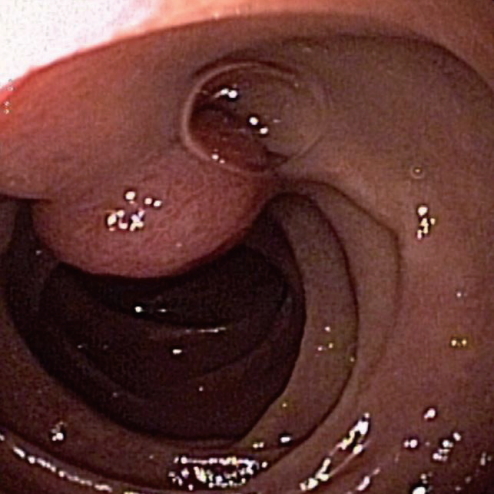
이에 측시경(side-viewing endoscope)을 이용하여 내시경 검사를 시행하였다. 십이지장 제2부의 바터 팽대부에 인접하여 게실이 보였고, 이에 연하여 약 2 cm 크기의 유경성 용종이 관찰되었다. 용종은 게실에 인접해 있었으나 바터 팽대부와는 분리된 병변이었다(Fig. 2A, 2B). 생검용 겸자를 이용하여 용종에서 조직생검을 시행하였고 병리 검사에서 만성 염증 소견이 보였다.

Fig. 2.

(A) Periampullary diverticulum and a pedunculated polyp were seen at examination with the side-viewing endoscope. (B) The ampulla of Vater (arrow) is observed. (C, D) The detachable snare was placed at the base of the stalk. (E) Polypectomy was performed with the snare (arrow: the detachable snare). (F) The residual neck (arrow) and the ampulla of Vater (arrowhead) are seen.

용종이 종양일 가능성을 완전히 배제할 수 없어 내시경 용종절제술을 시행하기로 하였다. 측시경을 삽입 후 용종 경부의 기저에 생리식염수와 에피네프린 혼합액을 주입하려 하였으나 겸자 올림장치의 굴곡으로 인해 주입기(injector)가 내시경 선단을 통과하지 못하여 점막하 주입(submucosal injection)을 시행할 수 없었다. 이에 박리성 올가미(detachable snare)로 용종 경부를 결찰 후(Fig. 2C, 2D) 올가미(snare)로 용종절제술을 시행하였다(Fig. 2E, 2F). 시술 후 출혈이나 천공 등 합병증은 발생하지 않았고 병리 검사 결과 과형성 용종(hyperplastic polyp)으로 확인되었다. 6개월 뒤 측시경으로 추적 관찰을 시행하였고, 잔여 병변은 관찰되지 않았다.

바터 팽대부는 십이지장 제2부의 주요 구조물로서 다양한 병변이 발생할 수 있어 상부위장관 내시경 검사 시 중요한 관찰 대상에 포함된다. 이를 위해 대한소화기내시경학회의 질 관리 프로그램에서도 상부위장관 내시경 검사 시 통상적으로 십이지장 제2부까지 관찰할 것을 권고하고 있다[1]. 하지만 직시경(forward-viewing endoscope)으로 바터 팽대부를 자세히 관찰하기에는 불리한 점이 많다. 우선 바터 팽대부가 직시경 하에서 비스듬하게 보이는 경우가 많고, 덮개주름(hooding fold)을 포함한 십이지장의 주름이나 유두 주위 게실에 의해 가려지는 경우도 있다[2]. 이전 연구 결과를 보면 직시경으로 바터 팽대부를 온전하게 관찰할 수 있는 경우가 24-54.7%에 불과하였고 34.6-45%에서는 바터 팽대부의 일부분이 관찰되었으며, 10.7-32%에서는 전혀 관찰할 수 없었다[3,4]. 바터 팽대부를 정확하게 관찰하기 위해서는 보통 측시경을 이용하며, 미국소화기내시경학회에서도 바터 팽대부 검사 시 측시경 사용을 추천하고 있다[5]. 하지만 국내에서 내시경을 시행하는 대다수의 의료기관이 측시경을 구비하고 있지 않아 검사에 어려움이 있을 것으로 예상된다. 최근 직시경에 투명캡을 씌워 관찰할 경우, 바터 팽대부의 관찰률을 높일 수 있다는 연구 결과가 보고되어 주목을 끌고 있다. 이 연구에서는 투명캡을 씌운 직시경을 이용할 경우, 바터 팽대부를 온전하게 관찰할 수 있는 경우가 90% 이상으로 보고되었다[2,4,6]. 따라서 측시경 검사가 여의치 않은 경우, 상황에 따라 직시경에 투명캡을 씌워 바터 팽대부 관찰을 시도해 볼 수 있다.

본 증례는 십이지장 용종이 직시경 검사에서는 바터 팽대부 종양으로 오인되었으나 측시경 검사 후 정확하게 진단된 경우로서, 바터 팽대부와 그 주변부의 병변을 평가할 때 측시경 검사의 중요성을 보여주는 좋은 사례이다.

References

1. Lee TH, Yoon JY, Paik CN, Choi HS, Jang JY. Updates on the facilities, procedures, and performance of the accredited endoscopy unit. Clin Endosc 2019;52:431–442.
2. Choi YR, Han JH, Cho YS, et al. Efficacy of cap-assisted endoscopy for routine examining the ampulla of Vater. World J Gastroenterol 2013;19:2037–2043.
3. Hew WY, Joo KR, Cha JM, et al. Feasibility of forward-viewing upper endoscopy for detection of the major duodenal papilla. Dig Dis Sci 2011;56:2895–2899.
4. Abdelhafez M, Phillip V, Hapfelmeier A, et al. Cap assisted upper endoscopy for examination of the major duodenal papilla: a randomized, blinded, controlled crossover study (CAPPA Study). Am J Gastroenterol 2017;112:725–733.
5. ASGE Standards of Practice Committee, Chathadi KV, Khashab MA, et al. The role of endoscopy in ampullary and duodenal adenomas. Gastrointest Endosc 2015;82:773–781.
6. Kallenberg FGJ, Bastiaansen BAJ, Dekker E. Cap-assisted forward-viewing endoscopy to visualize the ampulla of Vater and the duodenum in patients with familial adenomatous polyposis. Endoscopy 2017;49:181–185.

Article information Continued

Fig. 1.

Esophagogastroduodenoscopy revealed a duodenal mass, which masqueraded the tumor of the ampulla of Vater.

Fig. 2.

(A) Periampullary diverticulum and a pedunculated polyp were seen at examination with the side-viewing endoscope. (B) The ampulla of Vater (arrow) is observed. (C, D) The detachable snare was placed at the base of the stalk. (E) Polypectomy was performed with the snare (arrow: the detachable snare). (F) The residual neck (arrow) and the ampulla of Vater (arrowhead) are seen.