폐샘암종에서 원위총담관 전이로 인한 폐쇄성 황달
Obstructive Jaundice due to Metastasis from Pulmonary Adenocarcinoma to Distal Common Bile Duct
Article information

A 63-year-old woman presented with obstructive jaundice. Patient was diagnosed as stage III lung adenocarcinoma 3 years ago, and underwent surgery and concurrent chemoradiotherapy. Immunostaining revealed positivity for thyroid transcription factor-1 (TTF-1) and cytokeratin (CK) 7, and negativity for CK20. Fluorescent in situ hybridization test revealed tyrosine kinase inhibitor (ALK) gene rearrangement. However, right buccal metastasis with no other metastasis was found, and she had been followed up with palliative radiotherapy. Laboratory tests showed elevated total bilirubin (9.9 mg/dL [reference value, 0.2–1.2 mg/dL]). Abdominal computed tomography (CT) showed 2.4 cm ill-defined mass with abrupt narrowing at distal common bile duct (CBD) (Fig. 1A, arrow). Endoscopic retrograde cholangiography showed 2.5 cm-sized filling defect at distal CBD (Fig. 1B), and intraductal biopsy confirmed metastatic adenocarcinoma from lung cancer (Fig. 2A) with thyroid TTF-1 positivity (Fig. 2B). Other immunostaining results were positivity for CK7 and negativity for CK20. The patient underwent metallic stent insertion, and medication of crizotinib (ALK tyrosine kinase inhibitor). Malignant biliary tract obstructions of metastatic tumors could be originated from gastric, colon, breast and lung cancer. For lung cancers, most were small cell lung cancer which has an aggressive behavior [1], whereas metastasis from lung adenocarcinoma was rare. Although few cases of obstructive jaundice were caused by pancreatic or periampullary metastasis [2,3], CBD metastasis from lung adenocarcinoma was extremely rare. TTF-1 is known as positive in ~76% of lung adenocarcinomas. Accordingly, TTF-1 is important for differential diagnosis between primary cholangiocarcinoma and metastatic adenocarcinoma [4], being difficult with hematoxylin-eosin staining. Herein, we presented the case of distal CBD metastasis from lung adenocarcinoma harboring ALK gene translocation. TTF-1 expression is useful to make differential diagnosis from primary biliary tract cancer.
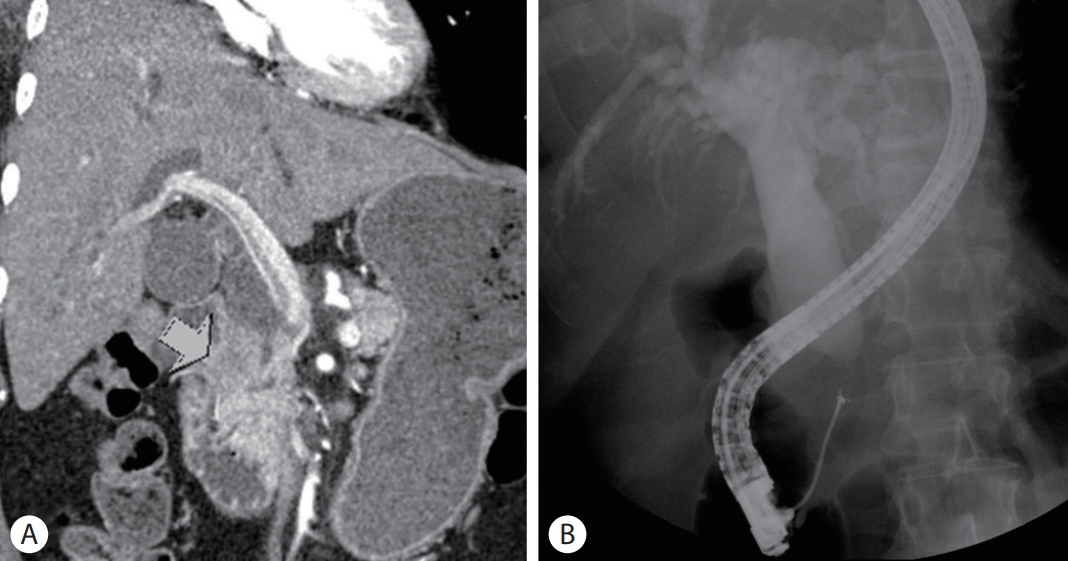
(A) Abdomen computed tomography showed biliary dilatation with abrupt narrowing at distal common bile duct. (B) Endoscopic retrograde cholangiography showed 2.5 cm-sized filling defect at distal common bile duct, and intraductal biopsy was performed.
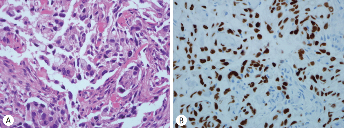
(A) Microscopic finding shows poorly differentiated adenocarcinoma (H&E, ×400). (B) Immunohistochemically, the tumor cells are positive for TTF-1 (H&E, ×400).