INTRODUCTION
Pancreatic pseudocyst is a common complication of acute or chronic pancreatitis, developing in up to 15% and 40% of these groups of patients[1]. Complications of pancreatic pseudocyst include infection, splenic vein thrombosis, rupture of pseudocyst, and massive hemorrhage into the pseudocyst[2]. Gastrointestinal (GI) bleeding is an uncommon but possible complication of pancreatic pseudocyst. This complication could occur after a fistula formation from the pseudocyst to the internal hollow organs[3]. Either upper or lower GI bleeding is possible according to the location of the fistula formation[4-6]. The gastric bleeding can be caused by the gastrocystic fistula formation and intracystic bleeding. Intracystic bleeding commonly originated from splenic artery pseudoaneurysm and this arterial bleeding could result in a fatal outcome[4,5]. Furthermore, there is a possibility of a massive upper GI bleeding, which is extremely rare[4,5].
Because GI bleeding associated with pseudocyst can be fatal, a spontaneous hemorrhage through gastrocystic fistula from the rupture of a pseudoaneurysm inside the pseudocyst should be suspected, in case the usual sources of bleeding are not detected through endoscopy. Here, we present a case of recurrent upper GI bleeding from gastrocystic fistula and pancreatic pseudocyst bleeding, which was controlled successfully by splenic artery embolization.
CASE
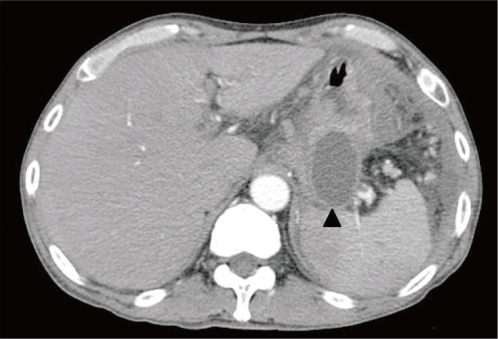
A 53-year-old male was referred to the emergency department due to melena and hematemesis. Melena has been presented for 2 days and a total amount of approximately 300 mL of blood from hematemesis was obtained at the time of referral. The patient’s past medical history revealed that he was a heavy alcohol drinker and has received treatment for alcoholic pancreatitis and pancreatic pseudocyst more than one month ago. His computed tomography (CT) scan, which he underwent during his past admission period, revealed a large pancreatic pseudocyst adjacent to the greater curvature side of his stomach with peripancreatic space infiltration and fluid collection (Fig. 1). However, the liver and spleen were relatively normal in appearance.
He had no previous history of peptic ulcer or gastrointestinal (GI) bleeding. The blood pressure of the patient in the emergency room was 86/60 mmHg and his pulse rate was 90 beats per minute. His conjunctivae were extremely anemic. His abdomen was relatively soft and flat. There was mild-tomoderate tenderness in the epigastric area. On laboratory examination, his hemoglobin was 6.9 g/dL, hematocrit was 20.6%, and other laboratory data were within normal range.
An emergency gastroduodenal endoscopic exam revealed a large fresh hematoma in the stomach without specific bleeding focus or current bleeding (Fig. 2). Three packs of red blood cells were transfused and the patient was transferred to the intensive care unit.
We performed a follow-up endoscopy after the stabilization of the patient to evaluate the bleeding focus. In the follow- up endoscopy, a large and extrinsic compression lesion was observed in the greater curvature side of the mid-tohigh part of the stomach. And an acute and large erosion was observed in the central area of the extrinsic compression lesion. At the time of the interval observation, a small amount of bloody fluid recurrently trickled out from the central erosion after saline irrigation (Fig. 3). This finding strongly suggested that the pancreatic pseudocyst abutted to the body of the extrinsically compressed stomach and caused a recurrent upper GI bleeding through the gastrocystic fistula from intracystic bleeding.
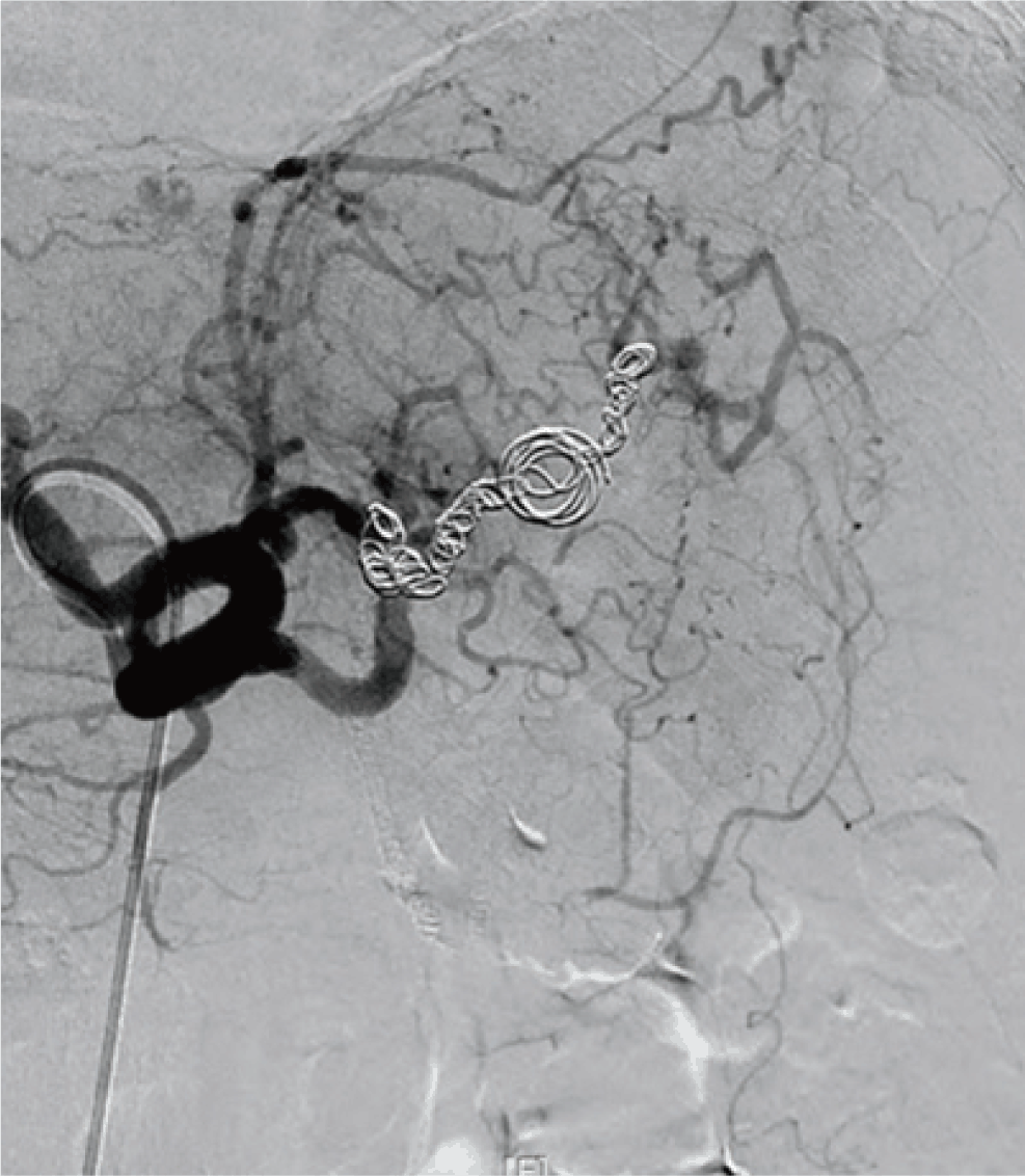
We immediately performed a contrast-enhanced CT scan to confirm the lesion. The CT scan revealed an intracystic hemorrhage of the pseudocyst which was located between the stomach and spleen. Furthermore, a splenic artery pseudoaneurysm was formed and an extravasation of the contrast media into the pseudocyst was also demonstrated (Fig. 4). Through these findings, we were able to confirm that the cause of the recurrent upper GI bleeding originated from gastrocystic fistula formation and intracystic aneurysmal bleeding. For the urgent angiography, the patient was transferred to Gachon University Gil Medical Center. Urgent angiography and therapeutic splenic artery embolization were successfully performed to stop further bleeding (Fig. 5). After the embolization, the patient’s clinical course was uneventful and no recurrence of bleeding has been reported after discharge.
DISCUSSION
A pancreatic pseudocyst is a cystic cavity contiguous with the pancreas and bound to it by epithelial inflammation. Unlike true cysts, a pancreatic pseudocyst lacks an epithelial lining and the epithelium of the cavity is usually lined with inflammatory tissues[7]. The most common cause of pancreatic pseudocyst is chronic alcohol abuse. Other causes are idiopathic, post operative, biliary tract disease, and abdominal trauma[8]. Signs and symptoms of pseudocyst are nonspecific which include abdominal pain, nausea, vomiting, weight loss, and epigastric tenderness. Ultrasonography and contrast-enhanced CT scan are most commonly used to establish the diagnosis of a pseudocyst[8]. Pancreatic pseudocyst can cause various complications which include obstructive jaundice, portal hypertension, infection, splenic vein thrombosis, rupture, and massive hemorrhage[2].
The bleeding complication of pancreatic pseudocyst in patients with chronic pancreatitis has been reported to be around 6-10%[9]. Serious bleeding into the gastrointestinal tract (stomach, duodenum and colon), retroperitoneum or peritoneum may occur. The overall mortality rate is approximately 13% in treated patients and 90% in untreated patients[9]. An upper GI bleeding from a pancreatic pseudocyst is a rare complication. Less than 1% of upper GI bleedings are caused by pancreatic pseudocyst[3]. The bleeding of a pancreatic pseudocyst commonly originated from pseudoaneurysm. The most common source of bleeding is the splenic artery (31%), followed by gastroduodenal (24%), superior and inferior pancreaticoduodenal (21%), and hepatic artery[10]. Three mechanisms of bleeding and rupture of the pseudocysts have been reported[11]. First, the inflammation and lytic enzymes of a pseudocyst can cause progressive digestion of the vessel wall. Therefore, erosion or disruption of the vessel wall is complicated. Second, the compression of a pseudocyst can produce ischemia of the vessel wall. Third, the inflammation or compression of a pseudocyst can cause thrombosis of the portal or splenic vein. Therefore, localized portal hypertension can be complicated.
In cases of a bleeding-complicated pseudocyst with or without pseudoaneurysms, several surgical procedures such as gastrectomy, splenectomy, bleeding arterial ligation, and pancreatectomy had been performed. The mortality rate was 33% following urgent operation[12]. Therefore, a surgical approach may be considered as relatively high risk. Another treatment option for a bleeding-complicated pseudocyst is angiographic embolization. A relevant data revealed that the success rate of angiographic embolization was 91% and the mortality rate was 14%[13]. An angiographic control of the bleeding was performed in other cases before undergoing elective surgery of a bleeding-complicated pseudocyst[14].
In this case, after the diagnosis of bleeding from a pancreatic pseudocyst, splenic artery angiography and angiographic embolization were successful. No further gastric bleeding was observed; therefore, a surgical approach was not performed. Even though angiographic embolization was successful in this case, we thought that the choice of a surgical procedure or angiographic embolization may vary in each patient’s clinical evolution.
In conclusion, we reported a case of a recurrent upper GI bleeding that originated from gastro-cystic fistula formation and intracystic aneurysmal bleeding of a pancreatic pseudocyst. And the massive gastric bleeding of a pancreatic pseudocyst is associated with high mortality. Therefore, an urgent contrast-enhanced CT scan with or without angiographic embolization or even surgery is necessary for proper diagnosis and treatment. The bleeding-complicated pseudocyst should be considered as one of the causes of recurrent GI bleeding in patients with pancreatic pseudocyst.


