INTRODUCTION
Acute pancreatitis is a reversible inflammatory disease of the pancreas that usually follows a mild and self-limited course. However, in approximately 15-20% of cases, acute pancreatitis progresses to serious adverse events, which may be life-threatening [1].
Gallstones and alcohol consumption are the leading causes of acute pancreatitis, accounting for 60-70% of cases. Other well-known causes of acute pancreatitis include post-endoscopic retrograde cholangiopancreatography (ERCP), abdominal trauma, post-operative state, adverse drug effects, sphincter of Oddi dysfunction, pancreatic cancer, periampullary diverticulum, pancreatic divisum and hereditary pancreatitis [2-4].
Although not common, hypercalcemia can cause acute pancreatitis if serum calcium levels increase rapidly. To date, few cases have been reported, all of which were self-limited and treated easily by conservative treatment including fasting, intravenous hydration, and removal of the predisposing factor [5-7].
Here we report a case of extensive necrotizing pancreatitis caused by excessive intravenous calcium supplementation.
CASE
A 26-year-old woman came to the emergency center of our hospital complaining of dizziness. Hours before the symptom started, she took 15 tablets of calcium channel blocker that she had mistaken for sleeping pills.
Her mental status was slightly drowsy, but orientations to time, person, and place were intact. At presentation, she had no gastrointestinal symptoms other than dizziness. Her initial blood pressure was 85/49 mmHg and heart rate was 101 beats/min. Initial laboratory data (normal range or value in parentheses) were as follows: white blood cell count, 9.4 × 10³/mm3 (4.0-10.0 × 10³/mm3); hemoglobin, 12.9 (12.0-16.0) g/dL; platelet count, 336 × 10³/mm3 (150-350 × 10³/mm3); corrected serum calcium 8.94 (8.6-10.2) mg/dL; phosphorus, 6.3 (2.5-4.5) mg/dL; blood urea nitrogen, 18 (10-26) mg/dL; creatinine, 2.13 (0.70-1.4) mg/dL; aspartate aminotransferase, 36 (0-40) IU/L, alanine aminotransferase, 21 (0-40) IU/L; alkaline phosphatase, 59 (40-120) IU/L; γ-glutamyl transferase, 105 (5-36) IU/L; total bilirubin, 1.3 (0.2-1.2) mg/dL; amylase, 40 (30-110) IU/L, and lipase, 17 (13-60) IU/L.
Normal saline hydration was used for volume resuscitation, and intravenous vasopressors (norepinephrine and epinephrine) were continuously infused to maintain blood pressure. In addition, glucagon, lipid emulsion and large doses of calcium compounds (calcium chloride and calcium gluconate) were intravenously administered during the initial 48 hours in the emergency center to treat calcium channel blocker intoxication [8]. Her vital signs stabilized and dizziness improved.
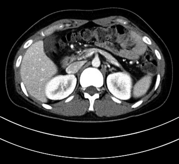
On day 3 after admission, the patient complained of sudden-onset severe epigastric pain and fever. Epigastric tenderness and decreased bowel sounds were noted on physical examination. Laboratory tests showed increased levels of serum pancreatic enzymes (amylase, 550 U/L; lipase, 1,679 U/L) and corrected serum calcium (17.24 mg/dL). Leukocytosis had developed in a neutrophil-dominant pattern in 94.6% of the total white blood cell count (19.4 × 10³/mm3). Abdominal computed tomography (CT) showed diffuse swelling of the pancreas with non-enhancing low attenuating regions, suggestive of necrotizing pancreatitis. Extensive peripancreatic fluid collection was also observed (Fig. 1). After being referred to the Department of Gastroenterology, she fasted and empirical broad-spectrum antibiotics were administered.
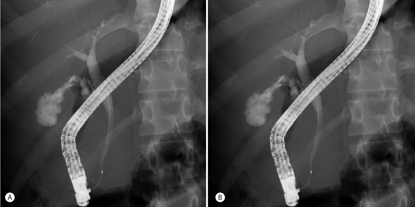
On day 11 after admission, she became icteric and her serum total bilirubin level increased to 4.3 mg/dL. ERCP was performed and a narrowed distal common bile duct was documented on cholangiography. A 10-Fr plastic stent was inserted into the narrowed common bile duct, following which her serum bilirubin level gradually decreased (Fig. 2).
On day 19 after admission, she complained of diffuse abdominal pain and fever. Follow-up CT showed walled-off necrosis and a large amount of peripancreatic fluid collection. Endoscopic ultrasonography (EUS) was performed using a linear echo-endoscope (GF-UCT260; Olympus Medical, Tokyo, Japan), and EUS-guided cystogastrostomy was performed using a partially covered self-expandable metal stent. After stent insertion, turbid fluid with necrotic debri gushed out through the stent. Fever and diffuse abdominal pain showed considerable improvement after the EUS-guided cystogastrostomy procedure (Fig. 3).
On day 22 after admission, abdominal pain of the right upper quadrant and fever recurred. Murphy’s sign was positive on physical examination. After CT, acute cholecystitis was highly suspected and percutaneous transhepatic gall-bladder drainage (PTGBD) was performed. Her symptoms were relieved after the PTGBD procedure (Fig. 4).
The patient eventually recovered well after supportive treatment and interventional procedures. The internal stents and PTGBD catheter were successfully removed within 3 months after their insertion. She was symptom-free and discharged 73 days after her admission. She has been regularly monitored at an outpatient clinic for the past 16 months since her discharge, and no additional events have been observed (Fig. 5).
DISCUSSION
Although not common, hypercalcemia can cause acute pancreatitis [9]. Mithöfer et al. [10] reported that acute pancreatitis occurred in rats after the administration of a calcium compound. As hypercalcemia developed, an increased in serum amylase and tissue trypsinogen activation peptide levels was observed along with edema formation and leukocytic infiltration in the pancreatic tissue.
The molecular mechanism underlying hypercalcemia-induced pancreatitis is yet to be elucidated. However, there are two suggested explanations. First, de novo activation of zymogens by hypercalcemia; the activated zymogens, including trypsin, destroy acinar cells and autodigest the pancreatic tissue, resulting in subsequent pancreatitis. Second, hypercalcemia can cause formation of pancreatic calculi and protein plug by modifying pancreatic secretion, resulting in pancreatic duct obstruction and subsequent pancreatitis. It has been suggested that other causes of acute pancreatitis, such as alcohol consumption, pancreatic ductal hypertension, ischemia, hyperlipidemia, and viral infection, also induce pancreatitis by increasing intracytoplasmic calcium levels [9].
There are many medical conditions known to cause pancreatitis by increasing serum calcium levels, including advanced malignancies with bone metastasis, multiple myeloma, vitamin D toxicity, sarcoidosis, and familial hypocalciuric hypercalcemia [9,11]. Although primary hyperparathyroidism is a well-known cause of hypercalcemia and the serum calcium levels of patients are usually high, the prevalence of acute pancreatitis among them is estimated to be only 1.5-7% because the serum calcium levels of these patients are maintained at consistently high levels above the normal range and do not rise abruptly [5]. Therefore, we can speculate that rapid increase in the serum calcium level is the key factor that causes acute pancreatitis. In our case, large doses of calcium compounds were administered to treat calcium channel blocker intoxication. Consequently, serum calcium level abruptly increased above 17 mg/dL, causing extensive necrotizing pancreatitis.
To the best of our knowledge, this is the first report of necrotizing pancreatitis caused by iatrogenic hypercalcemia. In this case, necrotizing pancreatitis could be life-threatening at presentation. However, the patient successfully recovered with supportive treatment and appropriate interventional procedures.
Although there are few reported cases of acute pancreatitis that developed as an adverse event of excessive calcium supplementation, all of them followed a self-limited course and improved with conservative treatment, including fasting and removal of the predisposing factor. Pronisceva et al. [12] reported a case of a 42-year-old woman complaining of vomiting and abdominal pain. She was diagnosed with acute pancreatitis, which was caused by hypercalcemia because of oral calcium supplementation. Calcium level in blood was returned to normal after cessation of calcium supplementation, and her symptoms were improved. Feyles et al. [13] reported a case of a 6-year-old boy with pseudohypoparathyroidism. He had been treated with calcium and vitamin D supplementation. He was admitted for acute pancreatitis, and his serum calcium level was high. Hypercalcemia induced by over-supplementation of calcium and vitamin D was thought to be the cause of his acute pancreatitis. His condition improved after calcium level in blood was returned to normal.
Physicians should learn from our case to pay special attention while administering calcium supplements for treating patients. Serum calcium levels must be periodically monitored and should be checked more frequently if intravenous calcium supplements are used. The possibility of acute pancreatitis and even necrotizing pancreatitis should be considered if the patient complains of abdominal pain and newly developed hypercalcemia. Severity should be assessed immediately, and appropriate management including interventional procedures should be considered.


