


INTRODUCTION
Although diffuse large B cell lymphoma (DLBCL) is the most common histologic subtype of non-Hodgkin lymphoma, a very few cases among them present with disseminated intravascular proliferation of lymphoid cells, which are categorized as intravascular large cell B-cell lymphoma (IVLBCL) [1,2]. IVLBCL is characterized by the proliferation of lymphoma cells within the lumen of small blood vessels, particularly capillaries and post-capillary venules without an obvious extravascular tumor mass or circulating malignant lymphoid cells in the peripheral blood [3]. Proper diagnosis of IVLBCL is still challenging since there are no pathognomonic clinical, laboratory and radiological findings. Herein, we describe a very unique case of 66-year-old female with IVLBCL found within a resected specimen of duodenal gastrointestinal stromal tumor (GIST).
CASE
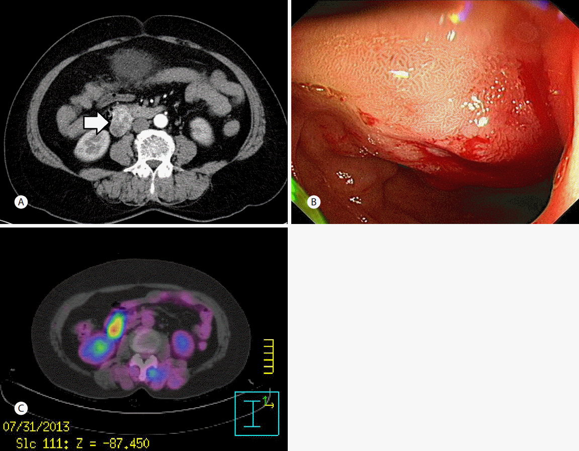
A 66-year-old woman was referred for further work-up of duodenal mass discovered incidentally on computed tomography (CT) scan. Two weeks earlier, she received CT scan for evaluation of chronic hematuria. Although the urological diagnosis was chronic cystitis, a large mass with heterogenous enhancement in the duodenum was found (Fig. 1A). Results of laboratory tests were as follows: white blood cell count, 6,290 µ/L (normal, 4,000 to 10,000 µ/L); red blood cell count, 4.60 × 10⁷ µ/L (normal, 4.0-5.5 × 10⁷ µ/L); hemoglobin concentration, 12.0 gd/L (normal, 12-16 g/dL); aspartate aminotransferase, 20 IU/L (normal, <37 IU/L); alanine aminotransferase, 16 IU/L (normal, <41 IU/L); alkaline phosphatase, 48 IU/L (normal 35-129 IU/L); lactic dehydrogenase, 233 IU/L (normal, 116-243 IU/L); C-reactive protein, 0.39 mg/dL (normal, <0.3 mg/dL).
Esophagogastroduodenoscopy was performed and a protruding mass with central umbilication was observed (Fig. 1B). Although the endoscopic impression was GIST, the result of biopsy was chronic inflammation with Brunner’s gland hyperplasia. On positron emission tomography (PET)-CT, the lesion showed highly increased metabolism with a maximal standard uptake value (SUV) of 10.6, which suggested high grade malignancy (Fig. 1C).
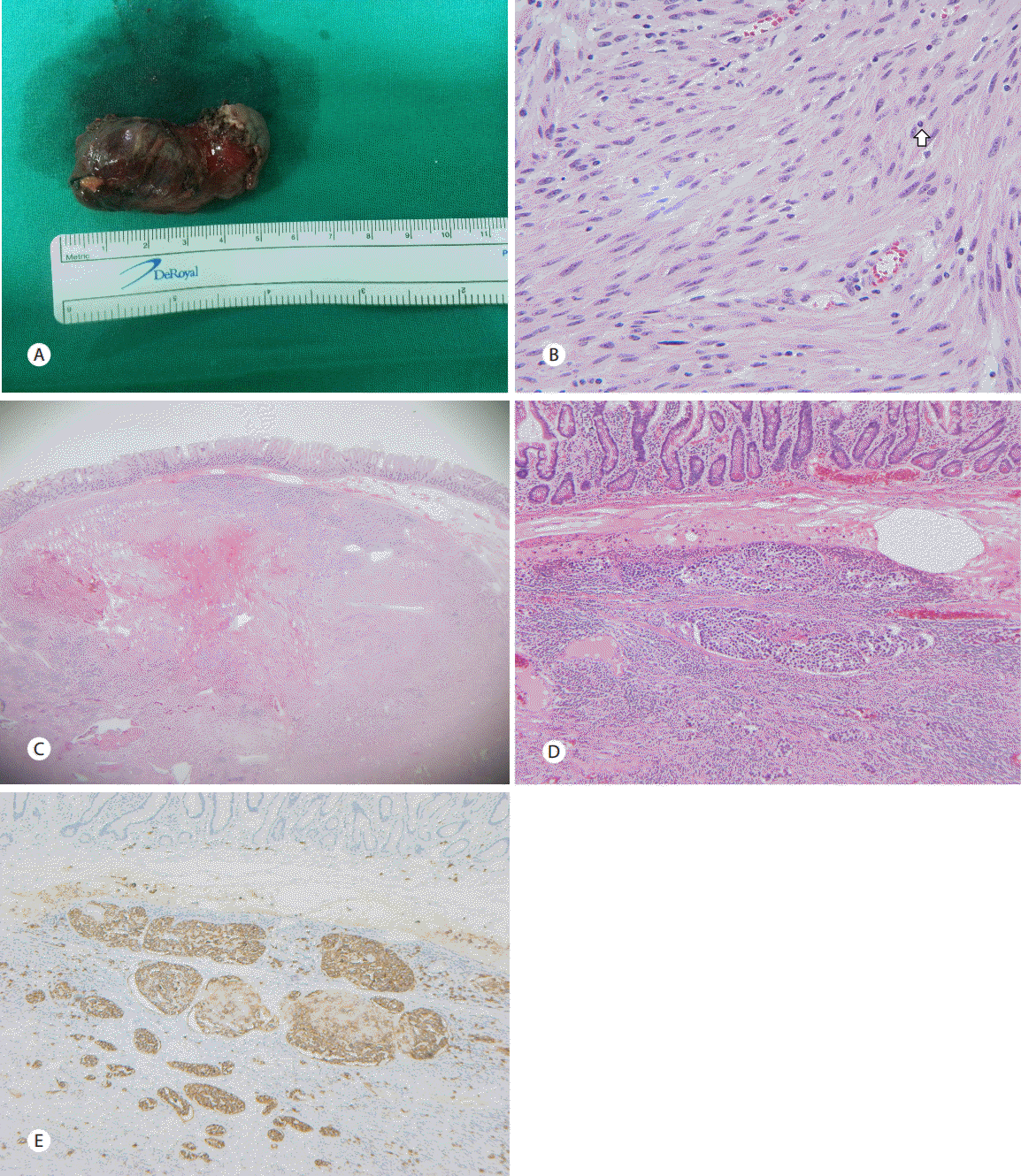
She received duodenal wedge resection with gastrojejonostomy. Gross pathology showed that the mass, measured 4.7 × 3.2 × 1 cm in size, was located mainly at antimesenteric side of the second to third portion of the duodenum (Fig. 2A). On microscopic examination, there were spindle cells with mild to moderate cellular atypia and the mitosis count was 3/50 high powered fields. Immunohistochemical staining for c-kit, smooth muscle actin and D2-40 were positive for the spindle cells. The resection margin was free from tumor cells and there was no invasion into the mucosa. Upon these findings, the patient was diagnosed with GIST of low risk of malignant potential by National Institute of Health, 2000 (Fig. 2B). Unexpectedly, however, there were nests of monotonous large nucleated cells within the submucosal vessels of the mass, which were positive for CD20 (Fig. 2C-E). There was no abnormal finding on bone marrow aspiration which was performed after the diagnosis of synchronous IVLBCL was made. The patient refused bone marrow study with additional chemotherapy and was lost to follow-up.
DISCUSSION
IVLBCL, a rare extranodal subtype of DLBCL, has poor prognosis due to its aggressive behavior and rapid systemic dissemination. More importantly, diagnosis is delayed frequently and most of the patients are diagnosed only when the disease has progressed. Although non-specific usually, there may be differences in the clinical characteristics according to the geographical origin of patients [4]. IVLBCL commonly involves the central nervous syndrome and skin in patients from the Western countries. Meanwhile, in asian patients, aggressive proliferation of normal histiocytes and T-lymphocytes resulting the hemophagocytic syndrome is predominantly accompanied [3]. Also, there are reports regarding the usefulness of laboratory values including pancytopenia, elevated lactate dehydrogenase, high serum betamicroglobulin, elevated Erythrocyte Sedimentation Rate, hypoalbuminemia [5]. However, these findings are non-specific and usually unhelpful for the diagnosis of IVLBCL. There was no suspicious clinical or laboratory abnormality in the present patient. Recently, there was a recent case report about the role of PET-CT to diagnose IVLBCL [6]. In this case also, the lesion showed highly increased metabolism with a maximal SUV value of 10.6. Although this finding suggested high grade malignancy [7-9], the malignant potential of GIST turned out to be low. Hence, it is possible that malignant cells of IVLBCL may have attributed to such a high maximal SUV [6,10].
In this case, IVLBCL was found in the surgical specimen of duodenal GIST, which is one of the infrequent primary gastrointestinal cancers comprising around. A recent data from Surveillance, Epidemiology, and End Results program of the National Cancer Institute had shown that the age-adjusted incidence of clinical GIST was 0.8 per 100,000 per year [11]. Also, diagnosis of IVLBCL in the GI tract is very unusual and there are just three cases reported in the English literature, one in the stomach [12], another in the ileum [13] and the other in the colon [14]. Therefore, co-existence of both tumors would be an extremely rare condition and, to the best of our knowledge, this is the first case report of IVLBCL with GIST in duodenum worldwide.
The initial therapy for IVLBCL is the combination of cyclophosphamide, doxorubicin, vincristine, and prednisone and anti CD-20 antibody, rituximab. Complete remission of IVLBCL is about 60% and about 3-year overall survival greater than 30% in current report [5]. In retrospective analysis of chemotherapy with or without rituximab, patients with chemotherapy plus rituximab had significant higher rates of complete remission, 2-year progression free survival, 2-year overall survival [3].
IVLBCL is a rare disease entity and difficult to diagnose due to its non-specific signs and symptoms but with poor prognosis without treatment. Due to lack of its specific diagnostic tool, incidental diagnosis and postmortem diagnosis is more common. In this case, we found IVLBCL with GIST initially with endoscopy and consequentially by surgical specimen. Although the patient was lost to follow-up in our hospital, diagnosis of IVLBCL changed the patient’s treatment plan in this case, so it is very important to diagnose IVLBCL accurately and in the process of diagnosis, clinician and pathologists’ role is essential. This is the first case of the coexisting IVLBCL and GIST reported in the world and extremely rare situation to our knowledge. It is critical to understand this rare disease entity and we need further research for the potentially useful diagnostic tools.