| Home | E-Submission | Sitemap | Contact us |  
top_img
Korean J Pancreas Biliary Tract > Volume 23(2):2018 > Article
담관낭종 불완전 절제 후 잔류 낭종에 발생한 거대 췌석
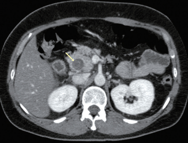
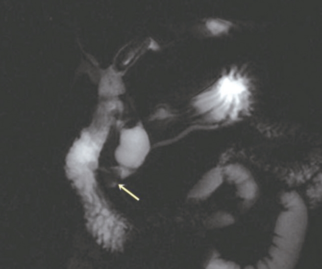
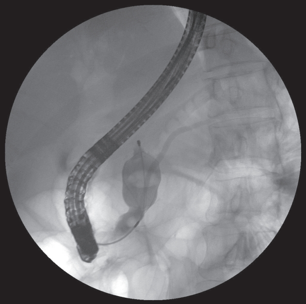
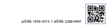
A 44-year-old female was admitted due to recurrent epigastric pain. She had undergone excision for a congenital choledochal cyst at the age of 27 years. Laboratory findings were as follows: white blood cell count of 4,600/mm3, total bilirubin of 0.7 mg/dL, aspartate transaminase of 38 IU/L, alanine transaminase of 20 IU/L, gamma-glutamyl transpeptidase of 30 IU/L, and serum amylase of 242 IU/L. Both abdominal computed tomography and magnetic resonance cholangiopancreatography showed findings compatible with remnant choledochal cyst with 15 mm sized pancreatic stone in the head portion (Fig. 1, 2). Endoscopic retrograde cholangiopancreatography revealed presence of anomalous pancreatobiliary junction, remnant choledochal cyst, and a filling defect in the pancreatic duct (Fig. 3). A white, fragile stone was removed after endoscopic sphincterotomy (Fig. 4). The patient remained asymptomatic without any complications after discharge. Although management of remnant choledochal cyst without symptoms is less clear [1,2], endoscopic sphincterotomy may be useful for the treatment and prevention of pancreatolith in patients with remnant choledochal cyst. Furthermore, complete excision of congenital choledochal cyst is necessary [3]. Incomplete resection of the cyst may cause pancreatic duct stone or protein plug formation in the remnant choledochal cyst [4,5].

REFERENCES

1. Matsuura K, Hashimoto D, Ikuta Y, Chikamoto A, Beppu T, Baba H. Education and imaging. Hepatobiliary and pancreatic: cystic bile duct remnant after surgery for a choledochal cyst. J Gastroenterol Hepatol 2013;28:754.
crossref pmid
2. Koshinaga T, Hoshino M, Inoue M, et al. Pancreatitis complicated with dilated choledochal remnant after congenital choledochal cyst excision. Pediatr Surg Int 2005;21:936-938.
crossref pmid
3. Ishibashi T, Kasahara K, Yasuda Y, Nagai H, Makino S, Kanazawa K. Malignant change in the biliary tract after excision of choledochal cyst. Br J Surg 1997;84:1687-1691.
crossref pmid
4. Nakano K, Mizuta A, Oohashi S, et al. Protein stone formation in an intrapancreatic remnant cyst after resection of a choledochal cyst. Pancreas 2003;26:405-407.
crossref pmid
5. Chiba K, Kamisawa T, Egawa N. Relapsing acute pancreatitis caused by protein plugs in a remnant choledochal cyst. J Hepatobiliary Pancreat Sci 2010;17:729-730.
crossref pmid

Fig. 1.
Abdominal computed tomography showed remnant choledochal cyst with a 15 mm sized hyperdense lesion in the pancreatic head portion (arrow).
kpba-23-2-87f1.jpg
Fig. 2.
Magnetic resonance cholangiopancreatography showed remnant choledochal cyst with a signal void lesion in the pancreatic head portion (arrow).
kpba-23-2-87f2.jpg
Fig. 3.
Pancreatic duct stone extraction was done by using a basket under fluoroscopic guidance.
kpba-23-2-87f3.jpg
Fig. 4.
Endoscopic view of the removed pancreatic duct stone. A white, fragile stone was removed after endoscopic sphincterotomy.
kpba-23-2-87f4.jpg
Editorial Office
Korean Pancreatobiliary Association #723 Le Meilleur Jongro Town, Jongro19, Jongro-gu, Seoul 03157, Korea
Tel: +82-2-2285-5145 Fax: +82-2-2285-5146   E-mail: kpba@kams.or.kr
About |  Browse Articles |  Current Issue |  For Authors and Reviewers
Copyright © 2023 by Korean Pancreatobiliary Association.     Developed in M2PI